
Fig. 1. A schematic illustration of the study design.
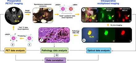
Ultrasmall fluorescent core-shell silica nanoparticles, cRGDY-PEG-CW800-C′ dots (1), were first labeled with iodine-124 (124I, t1/2 = 4.2 days) to form “hot” (or radioactive) 124I-cRGDY-PEG-CW800-C′ dots (2). A spontaneous melanoma miniswine was then peritumorally injected with (2) for detecting the metastatic disease based on whole-body PET/CT imaging (3). The lesion and the site of the SLN were confirmed and marked intraoperatively by a radiologist within the exposed surgical bed (4). Subsequently, serial injections of cold (or nonradioactive) αMSH-PEG-Cy5.5-C′ dots and cRGDY-PEG-CW800-C′ dots were performed peritumorally, followed by real-time multiplexed optical imaging using the Quest Spectrum imaging system (5) and resection of the nodes. Resected nodes then underwent ex vivo imaging (6) before histopathologic examination (7). Last, correlation analysis of the data derived from PET, optical, and pathological investigations was then performed (8). IHC, immunohistochemistry. (Photo credit: Feng Chen and Michelle S. Bradbury, Memorial Sloan Kettering Cancer Center)