Figure 6.
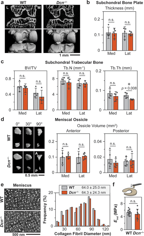
Decorin-null (Dcn−/−) murine knee joint does not show marked phenotype in the subchondral bone and meniscus. (a) Representative 2D μCT frontal plane images and reconstructed 3D images of the knee joint (L: lateral, M: medial). (b) Subchondral bone plate thickness and (c) subchondral trabecular bone structural parameters (BV/TV: bone volume/total volume, Tb.N: trabecular number, Tb.Th: trabecular thickness) of both medial and lateral tibia analyzed from μCT images. (d) Meniscal ossicles. Left panel: Representative reconstructed 3D μCT images (0°: top view, 90°: sagittal view) of meniscal ossicles (A: anterior, P: posterior). Right panel: Meniscal ossicle volume at anterior and posterior horns. (Panels b, c, d: mean ± 95% CI, n = 5). (e) Left panel: Representative TEM images of 3-month-old WT and Dcn−/− meniscus vertical cross sections. Right panel: Distributions of collagen fibril diameters are similar between the two genotypes (≥1000 fibrils from n ≥ 3 animals, p = 0.783). (f) AFM- nanoindentation (R ≈ 5 μm, nominal k ≈ 7.4 μm) yields similar modulus between WT and Dcn−/− meniscus surfaces (mean ± 95% CI, n ≥ 8, p = 0.335). Panels b–d, f: Each data point represents the average value measured from one animal.

