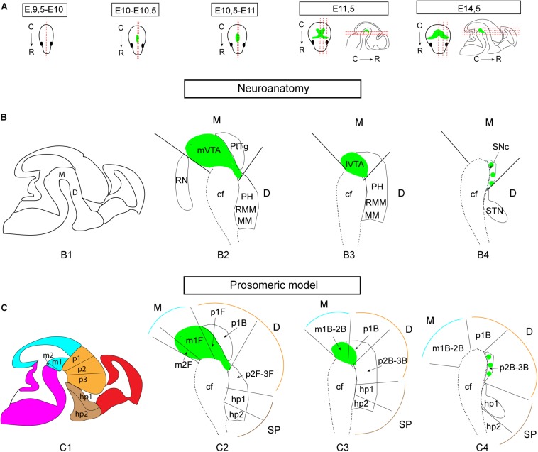
FIGURE 1.
Illustrations of the developing mouse brain to visualize sectioning angles and anatomical and neuromeric nomenclature implemented in the study. The midbrain DA system is shown in green for orientation, based on Th mRNA labeling. (A) Sagittal and horizontal sectioning angles of the brain at the different developmental stages used in the current study (E9.5-E10, E10-10.5, E10.5-E11, E11.5, and E14.5). (B,C) Schematic sagittal representations of three different medio-lateral levels of the brain of a E14.5 embryo (B1,C1) in which the mes-di-encephalic areas are shown in close-ups (B2–B4,C2–C4) to visualize the neuroanatomical (B) and prosomeric (C) terminology. The VTA is present in mesomers m1, m2, and prosomere (p) 1; the SNc is present in p2 and p3. Original drawings achieved by the use of published brain atlases (http://developingmouse.brain-map.org) and recent literature in which the medial section level is presented (Puelles and Rubenstein, 2003, 2015; Marín et al., 2005; Thompson et al., 2014; Puelles, 2019). Note that the VTA and SNc are part of the mesencephlon (M, blue) in standard anatomical terminology based on the columnar model, but part of both the M and diencephalon (D, orange) in the prosomeric model due to a change in the position of the border between M and D (Puelles, 2019). E, embryonic day; C, caudal; cf, cephahlic flexure; hp, hypothalamo-telencephalic prosomer; m, mesomer (mesencephalic prosomer); MM, mammillary nucleus; p, diencephalic prosomer; PH, posterior hypothalamus; PtTg, pretectal tegmentum; R, rostral; RMM, retromammillary nucleus; RN, raphe nucleus; SNc, substantia nigra pars compacta; STN, subthalamic nucleus; VTA, ventral tegmental area; SP, pallium/subpallium. (B) Refers to basal plate origin. F refers to floor plate origin.

