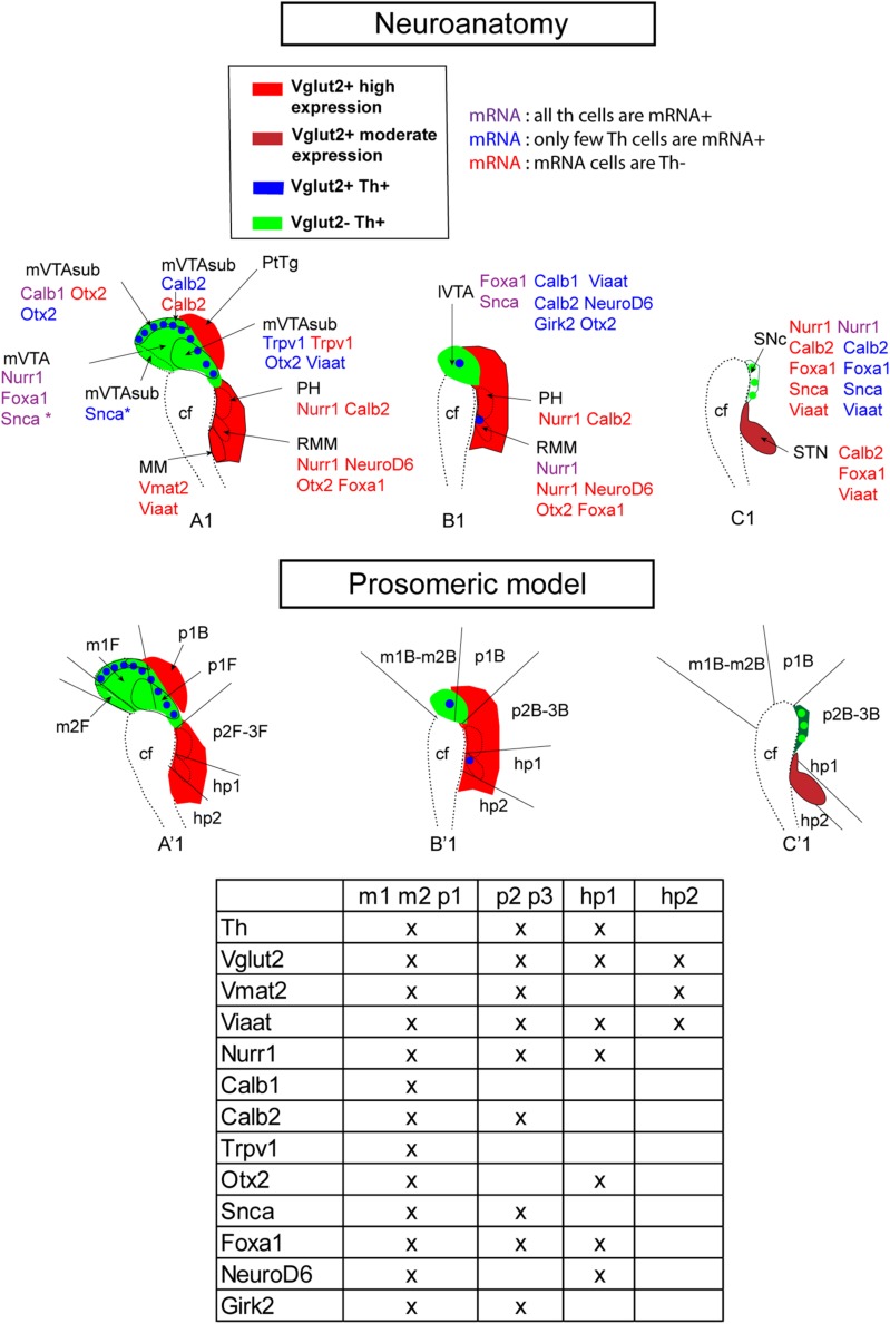
FIGURE 11.
Summary of current mRNA analysis presented in neuroanatomical (top) and prosomeric (bottom) terminology at E14.5. Sagittal sections of the mes-di-encephalic area in the developing mouse embryo at E14.5 illustrating representative section levels S1 (A1,A’1), S2 (B1,B’1), and S3 (C1,C’1) as indicated in Figure 1. Legend indicates color scheme for presence of Vglut2 and Vglut2/Th mRNAs. Note Vglut2 mRNA detection of moderate and high level indicated by different shades of red. Additional mRNAs, including vesicular transporter Viaat and Vmat2 (Figures 2, 3), and mRNAs selected for analysis based on presence in dopamine neuron subtypes (Figure 10) shown for each anatomical area (top panel) and prosomer identity (bottom panel, table). In top panel, each mRNA is presented in the context of its degree of co-labeling with Th mRNA (Th mRNA + or Th mRNA-). Caudal to the left; rostral to the right in each picture. cf, cepahlic flexure; hp, hypothalamo-telencephalic prosomer; m, mesomer (mesencephalic prosomer); MM; mammillary nucleus; p, diencephalic prosomer; PH, posterior hypothalamus; R, rostral; RMM, retromammillary nucleus; SNc, substantia nigra pars compacta; STN, subthalamic nucleus; VTA, ventral tegmental area; lVTA, lateral VTA; mVTA, medial VTA. (B) Refers to basal plate; F refers to floor plate.

