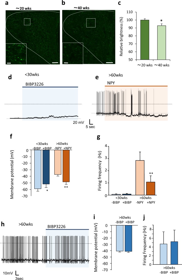
Figure 3.
Age-dependent activation of PVN neurons regulated by NPY in the PVN. (a–c), Confocal image of distribution of NPY-immunoreactive terminals/fibers in the PVN of rats aged around 20 weeks (18–23 weeks old) (a) and 40 weeks (39–44). Scale bars indicate 50 μm. (b) The bottom left square images are the enlarged images of the white square in each image. Scale bars in the enlarged images indicate 10 μm. Relative brightness acquired from image analysis of the confocal images (c). The brightness of NPY immunofluorescence in the PVN of rats aged around 20 weeks old was generalized as 100% (n = 8 each). *P < 0.05. unpaired t-test. (d) The representative recording of electrical activity of PVN neurons from rats aged <30 weeks after NPY antagonist (BIBP3226; 10−6 M) application. (e) Representative recording of electrical activity of PVN neurons from rats aged >60 weeks after NPY (10−8 M) application. (f) The membrane potential in the PVN neurons before and after application of BIBP3226 or NPY in rats aged <30 weeks and those aged >60 weeks, respectively (n = 6 each). *P < 0.05, **P < 0.01. paired t-test. (g) The firing frequency in the PVN neurons before and after application of BIBP3226 and NPY in the rats aged <30 weeks and those aged >60 weeks, respectively (n = 6 each). **P < 0.01. paired t-test. (h) The representative recording of electrical activity of the PVN neurons from the rats aged >60 weeks after NPY antagonist (BIBP3226; 10−6 M) application. (i–j) The membrane potential (i) and firing frequency (j) in the PVN neurons before and after application of BIBP3226 in the rats aged >60 weeks.

