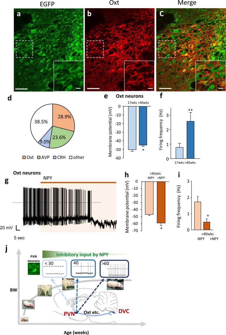
Figure 4.
Oxytocin neurons as one of the components of the PVN-DVC circuit. (a–c) The distribution of the EGFP-expressing neurons (a), Oxt-positive neurons (b) and merged image of a and b (c) in the PVN after DOX treatment continuously for two days. Scale bars indicate 50 μm. The image located in the bottom right is an enlarged image of the dotted square in each image, in which the scale bars indicate 10 μm. 3 V = third ventricle. (d) Percentage of Oxt, AVP, and CRH neurons among EGFP expressing neurons. (n = 3–4). (e) The membrane potential in PVN Oxt neurons at 17 weeks and >40 weeks. *P < 0.05. unpaired t-test. n = 29, 19, each. (f) The firing frequency in PVN Oxt neurons at 17 weeks and >40 weeks. **P < 0.01. unpaired t-test. n = 29, 19, each. (g), The representative recordings of the electrical activity of PVN Oxt neurons under NPY (10−8 M) treatment in the brain slices at >40 weeks. (h) The membrane potential in the PVN Oxt neurons before and after the application of NPY in rats aged >40 weeks. n = 6. *P < 0.05. paired t-test. (i) The firing frequency in the PVN Oxt neurons before and after application of NPY in rats aged >40 weeks. n = 6. *P < 0.05. paired t-test. (j) Summary of the mechanism to regulate age-dependent BW. BW increase continues until around 60 weeks. PVN neurons are relatively inactive in young rats (<30 weeks). Action potential firing and membrane potential increases in adulthood (around 40 weeks old) due to decreasing inhibitory inputs from NPY neurons with age. Upstream and downstream factors that regulate age-dependent activity of the PVN include NPY and Oxt, respectively. The PVN-DVC circuit is considered a regulator of age-dependent BW under NPY influence.

