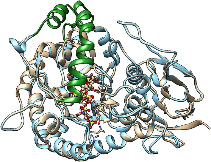
Figure 3.
Homology model of Hpa2 was built using the SWISS-MODEL server. PDB 5LA4 (Proheparanase) was used as a template. The obtained homology model of Hpa2 was then superimposed onto active heparanase (PDB code: 5e9c) cocrystal with heparin tetrasaccharide substrate. The linker (green ribbon) of Hpa2 partially occupies the heparin binding site. This is also true for proheparanase (PDB code:5LA4 structure). Heparanase is shown in golden, and Hpa2 in blue.

