Figure 4.
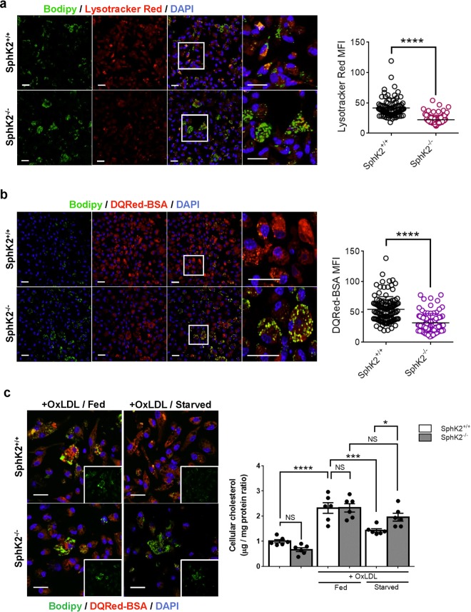
Impaired lysosomal activity in SphK2−/− macrophages. Peritoneal macrophages were freshly harvested from SphK2+/+ and SphK2−/− male mice after 2 weeks of WD feeding. (a) Staining of macrophages with the pH-sensitive LysoTracker Red and Bodipy. (b) Staining of macrophages with DQRed-BSA and Bodipy. (a,b) n = 5–6 mice per group. Total observed cell numbers were 88–117. Representative images (left). Scale bars, 20 μm. MFI of LysoTracker Red- and DQRed-BSA signals (right). (c) Effects of starvation on lipid deposition in macrophages. Peritoneal macrophages freshly harvested from non-WD fed mice were loaded with or without OxLDL for 4 h, followed by culture in normal growth medium (fed) or amino acid-depleted medium (starved) for 4 h (n = 6 per group). Representative images (left). Scale bars, 20 μm. Cellular cholesterol content (right). *P < 0.05, ***P < 0.001 and ****P < 0.0001. NS, not significant.

