Figure 1.
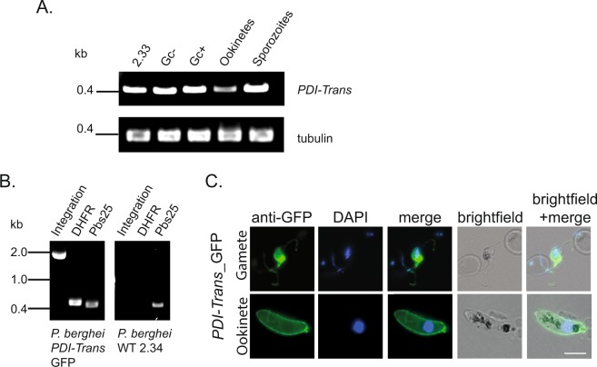
Constitutive expression of Plasmodium berghei PDI-Trans, and localization on the surface of gametocytes and ookinetes. (A) RT-PCR analysis of PDI-Trans in asexual blood stages using the non-gametocyte producing strain 2.33; non-activated (Gc-) and activated (Gc+) gametocytes; purified in vitro ookinetes and day 21 salivary gland dissected sporozoites. The analysis was complemented with alpha-tubulin loading controls (B). PCR confirmation of integration of egfp into the PDI-Trans locus. Oligonucleotides 35 and 14 were used to detect integration. Oligonucleotide 91 and 92 were used to detect DHFR presence, pbs25 oligonucleotides were used as positive controls. P. berghei WT 2.34 gDNA was used as a negative control for integration. (C) IFA of fixed, non-permeablised PDI-Trans-GFP parasites probed with anti-GFP; exflagellating male gametocytes (top) and ookinetes (bottom). Each panel shows an overlay of GFP fluorescence (green) and DNA labelled with DAPI (blue). White scale bar = 5 μm.

