Figure 5.
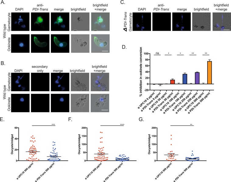
Anti-PDI-Trans antibodies inhibit fertilization and transmission in Plasmodium berghei. IFA of wildtype P. berghei ANKA male gametes and ookinetes with (A). anti PDI-Trans and (B). Secondary-only control antibodies (green) DAPI (blue). IFA of male gametes and ookinetes with anti PDI-Trans shows broad surface staining. White scale bars = 5 μm (C). IFA of ΔPDI-Trans gametes with anti PDI-Trans. Absence of staining illustrates anti PDI-Trans recognises native protein. White scale bar = 10 μm (D). Inhibition in ookinete conversion in in vitro ookinete development assay with anti PDI-Trans compared to negative control antibody UPC10 at concentrations of 0, 50, 100, 250 and 500 µg/ml. Asterisks indicate P value < 0.05 Paired t test, ns indicate P value not significant. (E–G) Triplicate standard membrane feeding assays with anti PDI-Trans compared with negative control antibody UPC10 at a concentration of 500 µg/ml. Individual data points represent the number of oocysts found in individual mosquitoes 12-days post feed. Horizontal bars indicate mean intensity of infection, whilst error bars indicate S.E.M within individual samples. Asterisks indicate P value < 0.05 Mann-Whitney U test, ns indicate P value not significant.

