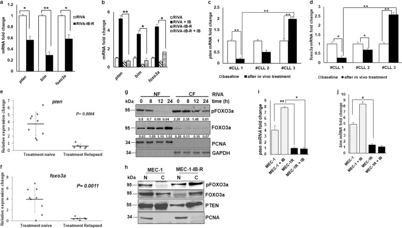
Fig. 3. Ibrutinib treatment regulates FOXO3a phosphorylation, nuclear translocation, and transcriptional activation of pten and bim.
a mRNA fold change of pten, bim, and foxo3a in parental vs IB-R RIVA cells after culture in the absence of ibrutinib for 72 h (*p < 0.05, **p < 0.01). SD is indicated as error bars (N = 3). b mRNA fold change of pten, bim and foxo3a in parental vs IB-R RIVA cells with or without ibrutinib (10 µM). c pten mRNA fold change was analyzed in primary cells obtained from 3 paired CLL patients’ samples pre- and post-ibrutinib treated in the clinic. SD is indicated as error bars (N = 1; triplicates of 1 experiment). d foxo3a mRNA fold change was analyzed in primary cells obtained from 3 paired CLL patients’ samples pre- and post-ibrutinib treated in the clinic. SD is indicated as error bars (N = 1, as in c). e, f relative expression changes in pten and foxo3a mRNA was analyzed in primary cells obtained from 9 treatment naive vs 5 treatment relapsed CLL patients’ samples after in vitro treatment with ibrutinib. Man–Whitney nonparametric analysis was performed to compare them. Two-sided p value for pten is 0.0004 and foxo3a is 0.0011. g Expression levels of pFOXO3a Ser253, FOXO3a, and PTEN in nuclear/ cytoplasmic fractions of parental RIVA cells after treatment with ibrutinib (10 µM) for the indicated time points. PCNA and GAPDH were used as a nuclear fraction and cytoplasm loading controls, respectively. h Expression levels of pFOXO3aSer253, FOXO3a, and PTEN in nuclear/cytoplasmic fractions of untreated parental and IB-R MEC-1 cells after culture in the absence of ibrutinib for 48 h. PCNA was used as a nuclear fraction loading control. MEC-1 parental and IB-R cells were treated with or without ibrutinib (10 µM) for 24 h followed by the ChIP assay. i, j qRT-PCR was performed for the pten and bim promoters. (*p < 0.05, **p < 0.01). SD is indicated as error bars (N = 2).

