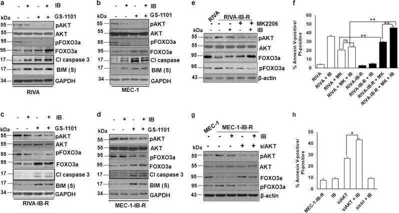
Fig. 5. PI3K/AKT inhibition upregulates FOXO3a levels and sensitizes IB-R cells.
a, b RIVA and MEC-1 cells were treated with ibrutinib (10 µM) and GS-1101 (5 µM) alone or in combination for 24 h. Expression levels of pAKT, AKT, pFOXO3aSer253, FOXO3a, cleaved Caspase 3 and BIM were determined by immunoblotting. GAPDH was used as a loading control. c, d RIVA-IB-R and MEC-1-IB-R cells were treated with ibrutinib (10 µM) and GS-1101 alone or in combination for 24 h. Expression levels of pAKT, AKT, pFOXO3aSer253, FOXO3a and cleaved Caspase 3 were determined by immunoblotting. GAPDH was used as a loading control. e RIVA-IB-R cells were treated with ibrutinib (10 µM) and AKTi (MK2206, MK) (5 µM for 48 h) alone or in combination. Expression levels of pAKT, AKT, pFOXO3aSer253, and FOXO3a were determined by immunoblotting. β-actin was used as a loading control. First lane indicates the baseline expression of proteins in parental RIVA cells. f RIVA and RIVA-IB-R cells were treated with MK2206 (5 µM) and ibrutinib (10 µM) either alone or in combination for 24 h. Cell viability was determined by Annexin V-PI staining. Control cells were treated with DMSO. (*p < 0.05). SD is indicated as error bars (N = 3). g MEC-1-IB-R were transfected with siAKT and treated with ibrutinib (10 µM) for 24 h. Expression levels of pAKT473, AKT, pFOXO3aSer253 and FOXO3a were determined by immunoblotting. β-actin was used as a loading control. First lane indicates the baseline expression of proteins in MEC-1 cells. h MEC-1-IB-R cells were transfected with siAKT or siControl and treated with ibrutinib (10 µM) for 24 h. Cell viability was determined by Annexin V-PI staining. Control cells were treated with DMSO. (*p < 0.05). SD is indicated as error bars (N = 3).

