Fig. 1.
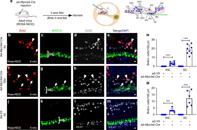
Myc and NICD co-activation induces proliferation in adult mouse cochlea in vivo. a A diagram illustrating the procedure of ad-Myc/ad-Cre injection in adult Rosa-NICD cochlea (left). A diagram depicts injection into the scala media (SM) of adult cochlea by cochleostomy (middle). Enlarged inset of a cross section shows cochlear structure and cell subtypes (right). Cld: Claudius’ cells; HeC: Hensen cells; OHC: outer hair cells; IHC: inner hair cells; IDC: interdental cells; DC; Deiters’ cells; OPC: outer pillar cells; TC: tunnel of Corti; IPC: inner pillar cells; IPh: inner phalangeal cells; NF: neuro fibers of auditory neurons; and IBC: inner cells. b–i Four (b–e) and 35 (f–i) days after injecting ad-Myc/ad-Cre mixture to the adult (6-week-old) Rosa-NICD cochleae in vivo, proliferating IHCs (MYO7A+/BrdU+, arrows) and SCs (SOX2+/BrdU+, arrowheads) were detected. j–m Four days after control ad-V5 in vivo injection into the adult Rosa-NICD cochleae, no proliferating cell was detected. n, o Quantification and comparison of number and percentage of BrdU+ IHCs and SCs in adult Rosa-NICD cochleae infected with ad-Myc/ad-Cre in vivo for four days (n) or 35 days (o) in the mid-base turn, compared to the ad-V5-injected adult Rosa-NICD cochleae. ***p < 0.001, ****p < 0.0001, two-tailed unpaired Student’s t-test. Error bar, mean ± s.d.; n = 8 for ad-Myc/ad-Cre groups, n = 6 for control groups in (n); n = 6 for each group in (o). n is the number of biologically independent samples. Scale bars: 10 μm. Source data are provided as a Source Data file.

