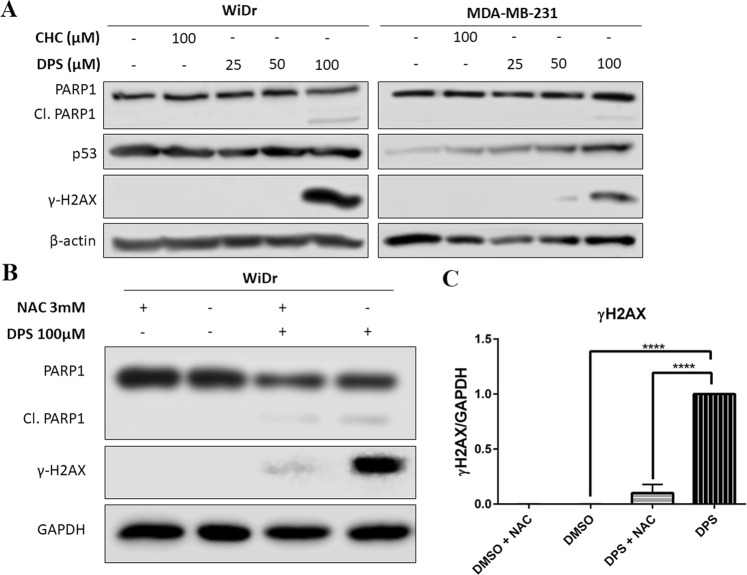
Figure 7.
(A) Treatment with 2a (DPS) for 24 hours induced PARP1 cleavage and histone H2AX phosphorylation in WiDr and MDA-MB-231 cells indicative of apoptosis and DNA damage. Further, treatment with 2a lead to an increase in p53 expression in MDA-MB-231 cells in a dose dependent manner. (B) Treatment with the radical scavenger N-acetyl cysteine (NAC) reversed H2AX phosphorylation but not PARP1 cleavage in WiDr cells. (C) Densitometry analysis of γ-H2AX when compared to GAPDH as a loading control. Representative western blots of three independent experiments, and are cropped from the full-length images. Full-length blots can be found in the supplementary information, Figs. S2–S4. Repeated measures one-way ANOVA was used to calculate statistical significance (****P < 0.0001).

