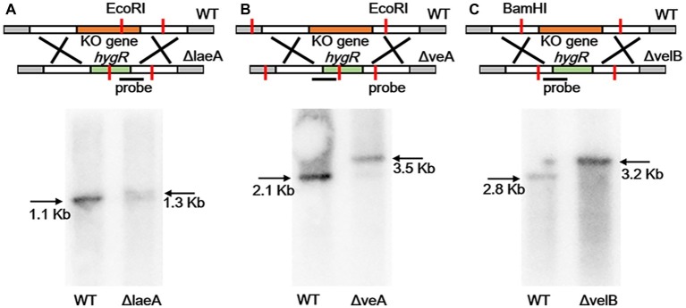
Figure 2.
Southern blotting verification of laeA, veA, and velB gene deletion. (A) The WT and ΔlaeA isolates were digested with EcoRI. A fragment amplified from ΔlaeA was used as the probe. (B) The WT and ΔveA isolates were digested with EcoRI. A fragment amplified from ΔveA was used as the probe. (C) The WT and ΔvelB isolates were digested with BamHI. A fragment amplified from ΔvelB was used as the probe.

