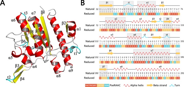
Figure 1.

A schematic view of a protein 5TCD in PDB with secondary structures. Subfigure (A) shows the three-dimensional structure of this protein. All secondary structural elements are indicated as different labels. Subfigure (B) shows its corresponding chain view, where the gray background represents the portion of the reduced amino acid sequence that matches the protein secondary structural elements.
