
Fig. 7.
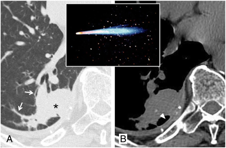
Comet tail sign. Axial CT image (a) shows a round consolidation (black asterisk) and stretched vessel and bronchus (white arrows): these combinations—reproducing a “comet tail” at the border of a round lesion—suggest the presence of round atelectasis. On axial CT image (b, mediastinal window), a dorsal pleural plaque with calcification is also appreciable (white arrowhead)