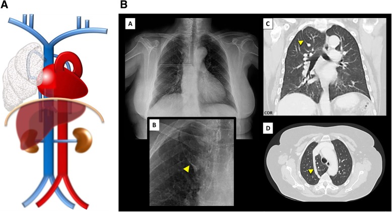
Fig. 14.
a Azygos fissure and lobe schematic. Resulting from an abnormal course of the azygos vein in the apex of the right lung subsequent to a failure of migration. b Azygos fissure and lobe imaging. Azygos fissure and lobe, as seen on chest X-ray [A, B—magnification view], as a line that crosses the apex of the right lung ending in a tear shape, and on thoracic CT scan on coronal [C] and axial [D] planes

