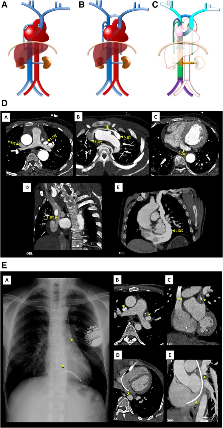
Fig. 3.

a Left SVC schematic. A single left SVC, with its usual course lateral to the aortic arch and drainage into the coronary sinus. b Double SVC schematic. The most frequent case of left SVC persistence, in which there are two SVC, each running on one side of the mediastinum, the right draining normally and the left one draining typically to the coronary sinus. These may or, more frequently, may not be connected by the left brachiocephalic vein. c Double SVC embryology schematic. Double SVC resulting from the persistence of the left anterior cardinal vein, here connected by the inter-anterior cardinal anastomosis (dark blue), precursor of the left brachiocephalic vein. If, in addition, there was regression of the right anterior cardinal vein, the end result would be a single left SVC. d Double SVC imaging. Contrast-enhanced CT showing double SVC, with a left-sided SVC (L-SVC) along the left side of the mediastinum [A] and draining into an enlarged coronary sinus [C–E], connected to a normal right-sided SVC (R-SVC) by the left brachiocephalic vein (asterisk) [B]. e Persistent left SVC eventuated by left-sided implantable cardioverter defibrillator leads. Chest X-ray [A] and contrast-enhanced CT scan [B–D] showing persistent left SVC (triangle) eventuated by a left-sided implantable cardioverter defibrillator lead, entering the right atrium via the coronary sinus (arrow)
