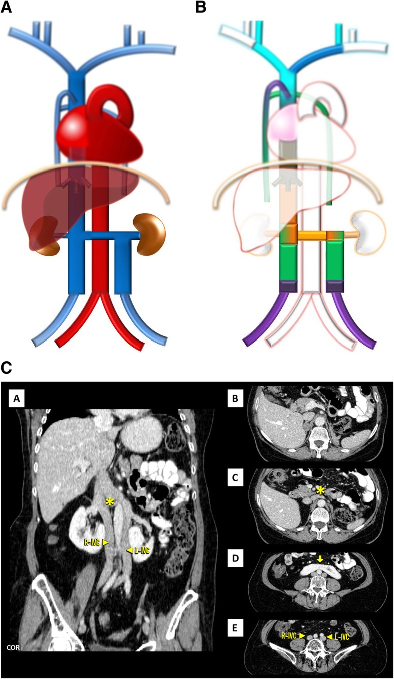
Fig. 6.

a Double IVC schematic. With the left component draining to the left renal vein, which joins the right IVC as normal. b Double IVC embryology schematic. Double IVC resulting from the persistence of both supracardinal veins, uniting in a single trunk on the right through the sub-supra and inter-subcardinal anastomosis, forming the left renal vein. c Double IVC imaging. Contrast-enhanced CT showing duplicated IVC, with a normal right-sided IVC (R-IVC) and a left-sided IVC (L-IVC) which ends at the left renal vein (asterisk) [C], note also horseshoe kidney (arrow) [D]
