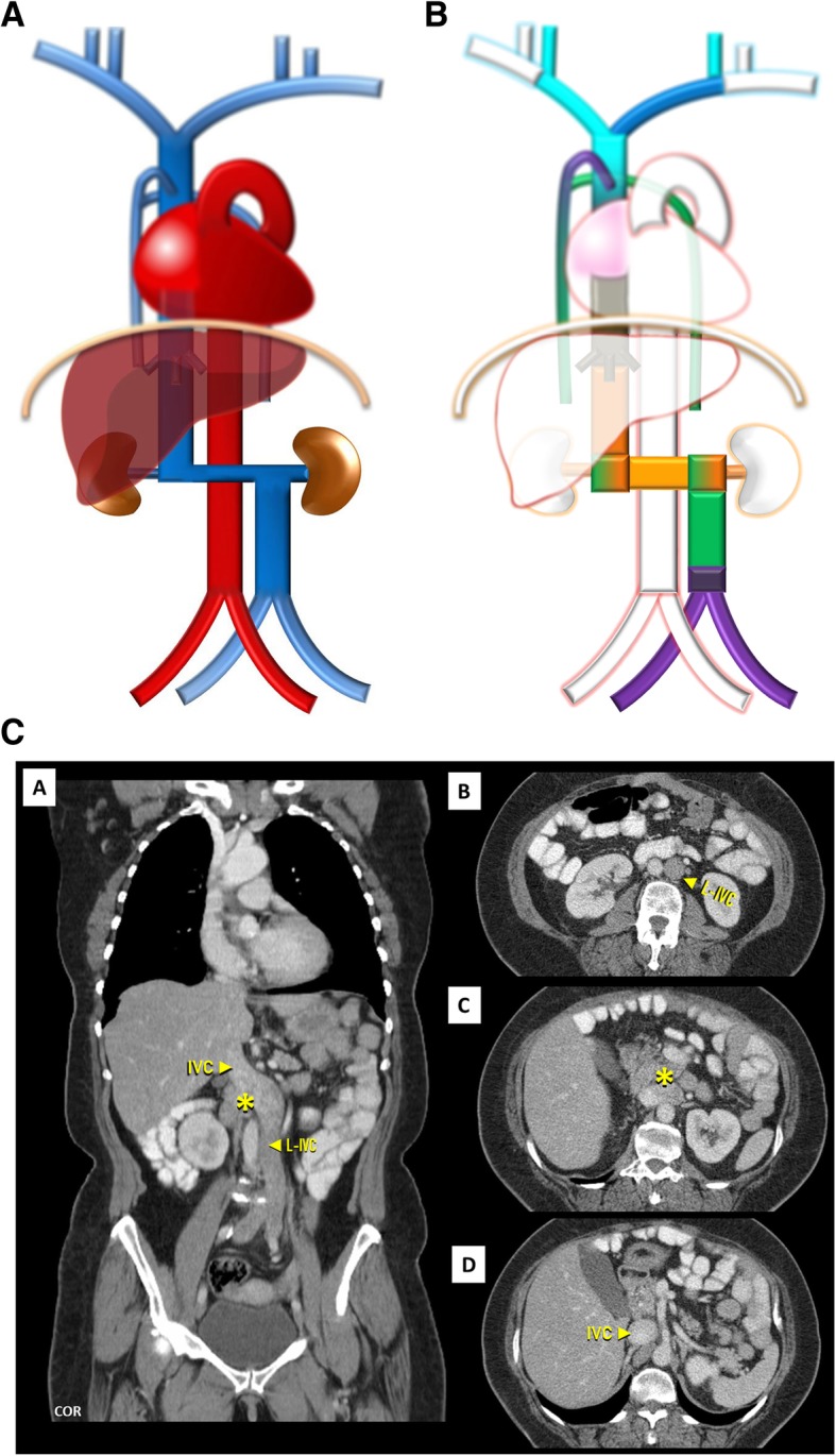
Fig. 7.

a Left-sided IVC schematic. Being different from duplication as the infrarenal segment of the right IVC is absent .The left IVC joins the left renal vein, which in normal fashion crosses ventrally the aorta to join the right renal vein from which results a normal suprarenal segment of the IVC. b Left-sided IVC embryology schematic. Left-sided IVC resulting from the regression of the right supracardinal vein, in addition to the persistence of the left supracardinal vein. c Left-sided IVC imaging. Contrast-enhanced CT showing a left-sided IVC (L-IVC), which characteristically joins the left renal vein, who in a normal fashion crosses ventrally to join the right renal vein (asterisk) and form a normal suprarenal segment of the IVC
