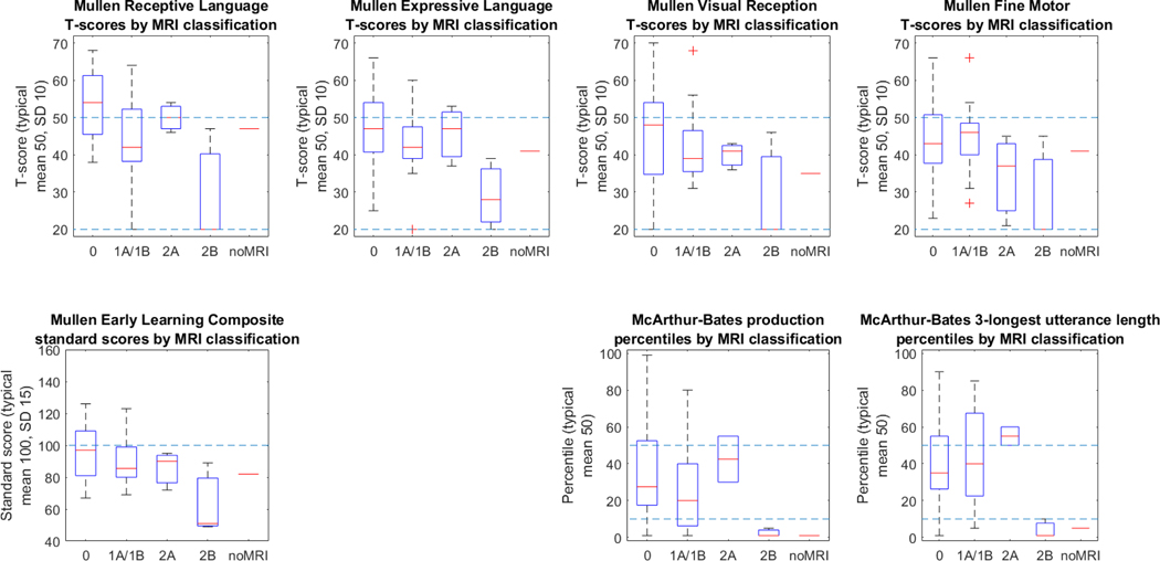
Figure 2: Assessment outcomes by modified NICHD MRI grade.

NICHD MRI grade reflects both the degree and gross distribution of brain injury on conventional MRI. Grades include categories for normal MRI (0), cortical/subcortical involvement only (1A (minimal cerebral lesions) and 1B (more extensive cerebral lesions), basal ganglia/thalamic/deep white matter involvement only (2A (basal ganglia, thalamic, internal capsule lesions only), or involvement of both areas (2B (basal ganglia, thalamic, internal capsule, and cerebral lesions) and 3 (cerebral devastation)). T-scores are normed against typical validation groups with mean scores of 50 (higher dotted line) and a standard deviation of 10; a T-score of 20 represents the floor of the instrument for individual subscores (lower dotted line).
