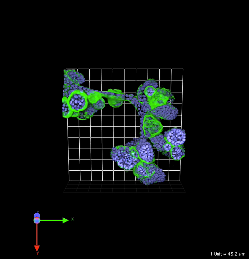
Fig. 1.
MCF-10A human breast epithelial cells develop over time into clusters of acini linked by tubules when grown in 3D rBM overlay monocultures. Image is 3D reconstruction of optical sections taken through entire volume on a Zeiss LSM-510 META confocal microscope. Structures were fixed and processed at 21 days of culture and stained for human laminin 332 to indicate production of human basement membrane proteins in these cultures (green) and Hoechst 33342 as a marker of nuclei (blue). One grid is 45 μm

