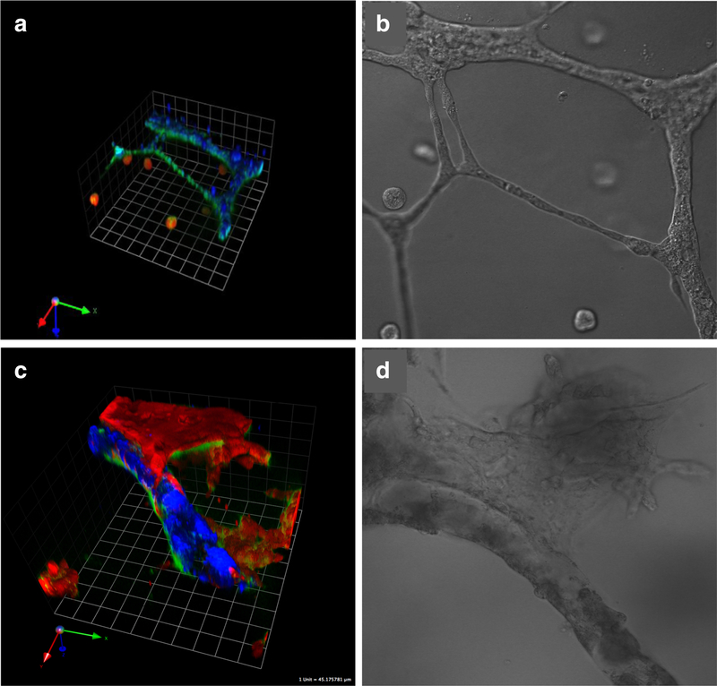
Fig. 5.
Human DCIS cells migrate toward the tubular networks formed by human umbilical vein endothelial cells over time in 3D rBM overlay co-cultures, attach and intravasate into the tubules. In movies of 3D reconstructions of optical sections, individual DCIS cells can be seen within the tubules. There is in parallel a striking increase in the growth and invasive phenotype of DCIS structures from 2 days in culture (a, b) to 5 days (c, d). DCIS cells were transduced with LentiRFP (red) and endothelial cells labeled with CellTrace Far Red (pseudocolored blue). Green fluorescence represents degradation products of DQ-collagen IV. Images of live co-cultures were obtained on a Zeiss LSM-510 META confocal microscope and confocal image stacks reconstructed in 3D with Volocity software. One grid is 45 μm

