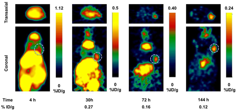
Fig 3.
PET images showing transaxial (top) and coronal (bottom) slices of mice bearing MCF7 tumor xenografts (s.c., dotted white circle) on right forelimb administered with [124I]IPAG at 4, 30, 72 and 144 h time points. It is clear from the images that the activity clears from the circulation at later time points providing high contrast images of the tumor.

