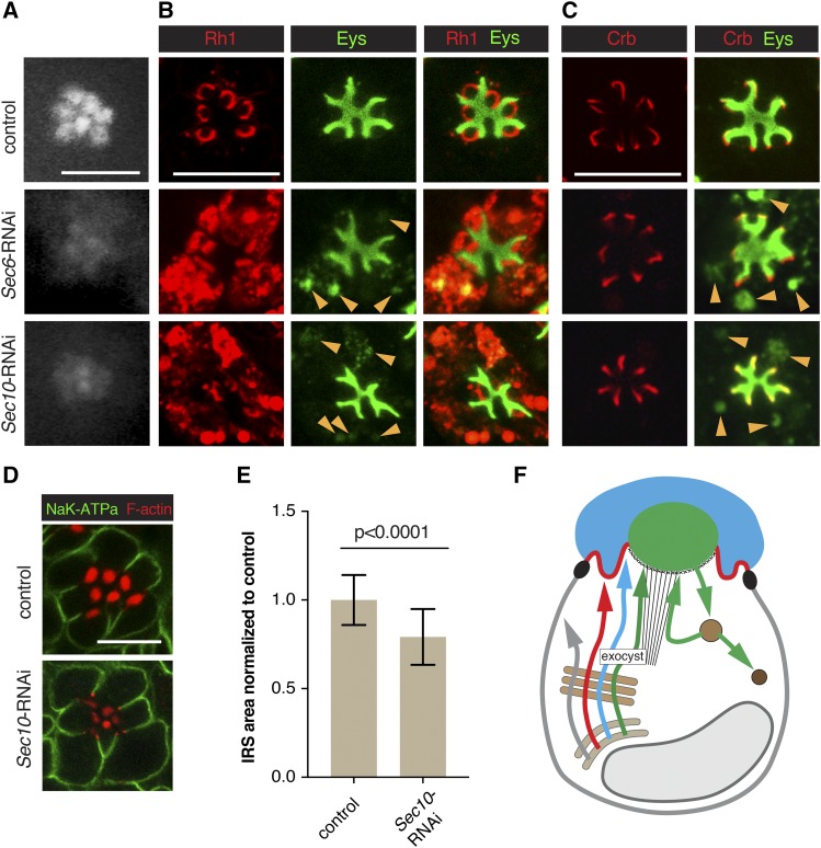
Figure 4.
The exocyst contributes to Rh1 and Eys exocytosis. RNAi line VDRC 22079 (Sec6-RNAi) was used to knock down Sec6, and line BDSC 27483 (Sec10-RNAi) to deplete Sec10. Sec6-RNAi or Sec10-RNAi were crossed with UAS-dicer-2; pGMR-Gal4. UAS-dicer-2/+; pGMR-Gal4/+ was used as control. Scale bars, 5 μm. (A) Individual rhabdomeres were only partially visible in Sec6 and Sec10 deficient retinas using TLI. Both were categorized as Class I. (B) Sec6 and Sec10 deficient PRCs show cytoplasmic accumulation of Rh1 and Eys (arrowsheads). Cytoplasmic Eys colocalizes with Rh1. (C) No defect was observed for Crb levels or localization in exocyst knockdown PRCs. Cytoplasmic Eys in exocyst deficient PRCs is visible in the absence of Rh1 staining (arrowheads), eliminating the possibility of cross-reaction between Eys and Rh1 antibodies as an explanation for the cytoplasmic Eys signal seen in (B). (D) Levels/localization of the basolateral protein Nrv (K+Na+ATPase subunit) was not affected in exocyst compromised PRCs. Acti-stain555 (F-actin) was used to visualize the rhabdomeres. (E) IRS size was significantly reduced in Sec10 deficient retinas. A total of 69 individual IRS were measured for control and 118 for Sec10 RNAi ommatidia, using three different animals per genotype. Values were normalized to the control. Error bars represent standard deviation. Unpaired non-parametric Mann-Whitney test. (F) Summary model indicating the secretion of Rh1 and Eys containing secretory vesicles depends on the exocyst. See Figure 1A for annotation and text for discussion.

