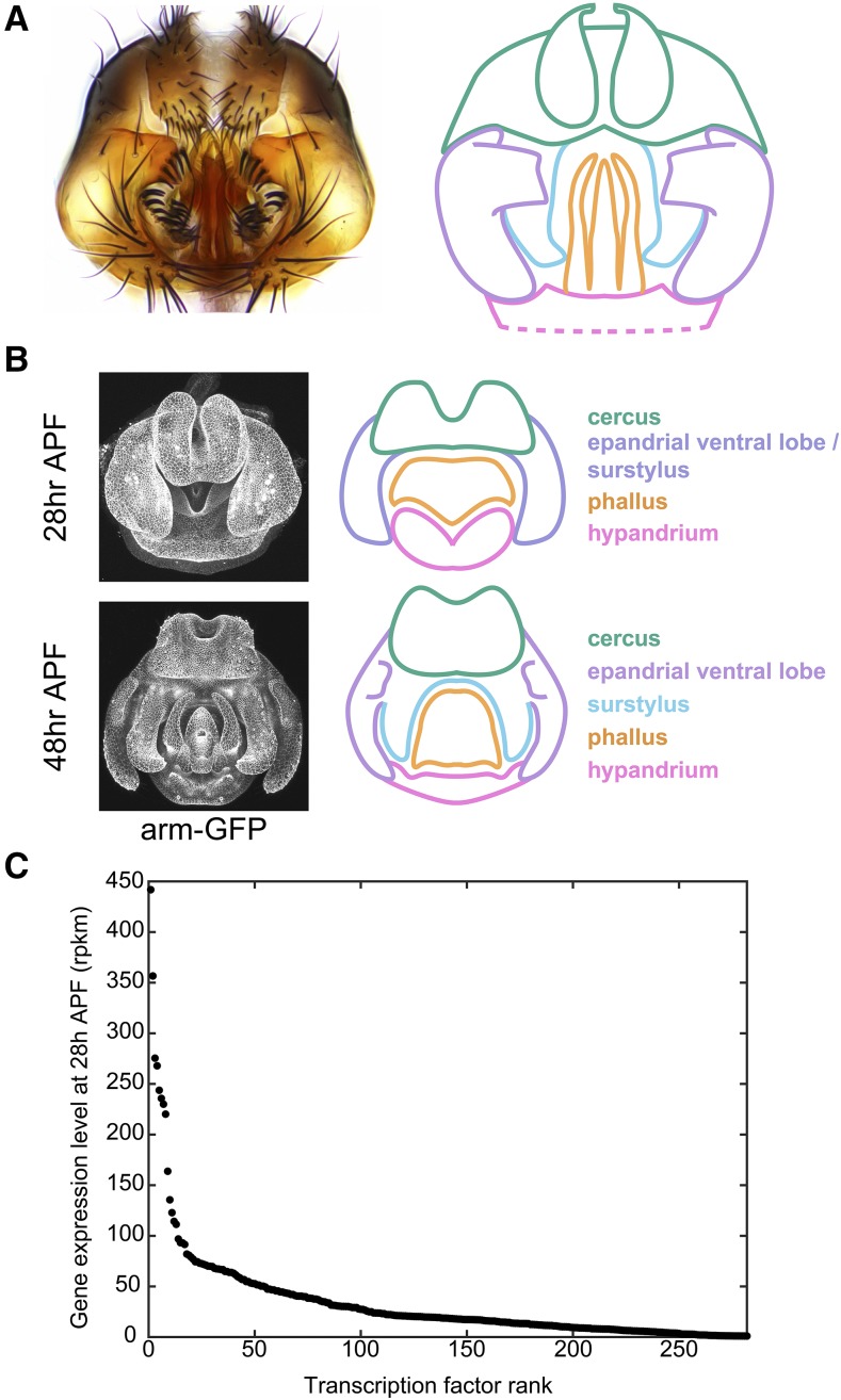
Figure 1.
Overview of male terminalia in Drosophila melanogaster. A) Left: light microscopy image of adult male terminalia. Right: schematic of major terminal structures. Pink: hypandrium; orange: phallus; light purple: epandrial ventral lobe; cyan: surstylus; green: cercus. The hypandrium extends beyond the cartoon, as represented by dotted lines. Note that our annotation of the cercus includes epandrial dorsal lobe (EDL) and subepandrial sclerite; these are difficult to distinguish during development and thus have been collapsed under the umbrella of cercus structures. B) Left: confocal microscopy images of developing male terminalia at two developmental time points in a transgenic line where apical cell junctions are fluorescently labeled using an armadillo-GFP fusion transgene. Right: schematic of major terminal structures in development, color coded as above. Dorsal-ventral (D-V), and medio-lateral (M-L) axes are labeled. Anterior structures project into the page, while the posterior end projects out of the page. C) Expression levels in reads per kilobase per million mapped reads (rpkm) of the 100 most highly-expressed transcription factors at 28 hr after puparium formation (APF) as measured by RNA-seq.

