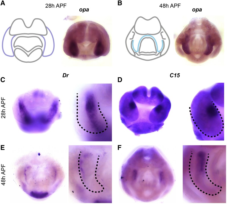
Figure 3.
Transcription factors expressed in the surstylus. A) Left: schematic of major terminal structures at 28 hr APF with the epandrial ventral lobe / surstylus indicated in dark purple. Right: Light microscopy image of in situ hybridization data for odd paired (opa) mRNA at 28 hr APF. B) Left: schematic of major terminal structures at 48 hr APF with the surstylus outlined in cyan. Right: Light microscopy image of in situ hybridization data for opa mRNA at 48 hr APF. (C,E) in situ hybridization data for Drop Dr mRNA in whole terminalia (left) and at higher magnification (right) at 28 hr APF (C) and at 48 hr APF (E). (D,F) in situ hybridization data for C15 mRNA in whole terminalia (left) and at higher magnification (right) at 28 hr APF (D) and 48 hr APF (F). Dashed lines indicate the boundary of the EVL/surstylus (C and D) or the surstylus (E and F).

