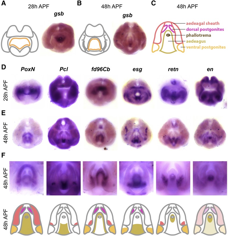
Figure 6.
Transcription factors expressed in the phallus. A) Left: schematic of major terminal structures at 28 hr APF with the phallus indicated in orange. Right: Light microscope image of in situ hybridization data gooseberry gsb mRNA at 28 hr APF. B) Left: schematic of major terminal structures at 48 hr APF with the phallus indicated in orange. Right: Light microscopy image of in situ hybridization data for (gsb) mRNA at 48 hr APF. C) Cartoon representation of the substructures of the phallus: ventral postgonite (orange), aedeagus (yellow), phallotrema (brown), dorsal postgonites (pink), and aedeagal sheath (red). Additional in situ hybridization data for transcription factors Poxn, esg, fd96Cb, retn, Pcl, and en at 28 hr APF (D) and 48 hr APF (E). F) Top: High magnification images of the samples shown in (E) to illustrate details of phallus expression patterns. Bottom: Cartoon representation of the substructures of the phallus, with shading indicating expression within each substructure. Note that for en, light shading indicates weak expression throughout the phallus.

