Figure 3.

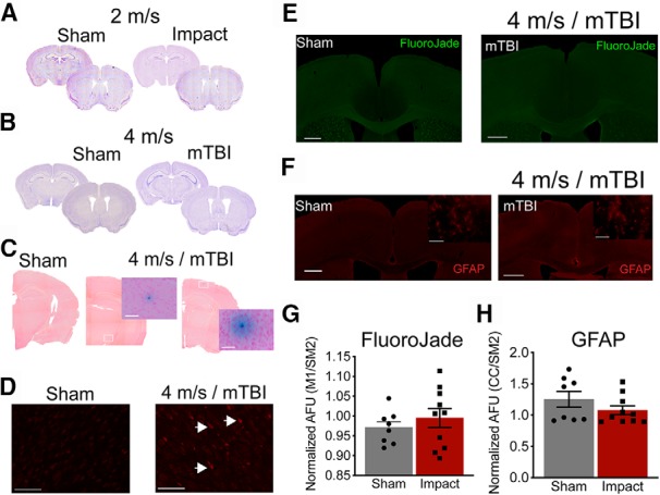
Impacts at 4 m/s (mTBI) do not produce significant tissue damage or astrocyte activation. Representative Nissl stains indicating no gross structural damage 24 h post-impact for 2 m/s (A) or 4 m/s (B). Prussian blue stain was used to identify microbleeds and the tissue was counter stained with NucRed. Three to four sections per animal were stained, and we only identified the two microbleeds shown in C from two separate mTBI animals. No other microbleeds were present in the sham or the other six mTBI animals. Representative images for sham-treated animals and the two microbleed sections taken at 4× and 40× magnification images of the microbleed themselves (C). The TUNEL stain was used to identify cells undergoing programed cell death. We only identified one mTBI animal that had detectable staining. Representative images are shown for sham-treated animals and the one mTBI animal that had TUNEL-positive cells shown by the arrow heads (D). Cell death was also assessed using FluoroJade-C. We were unable to detect a difference in overall fluorescent or individual cell bodies with positive staining. Representative images are shown (E). Neuroinflammation was assessed by GFAP staining. Representative images are shown (F). Again, we were unable to detect a difference in the overall fluorescence between the sham or mTBI (4 m/s) animals. Quantified fluorescence was measured by averaging the pixel intensity of the dorsal motor cortex to the lateral somatosensory cortex for sham and mTBI animals for FluoroJade (G) and GFAP staining (H). Scale bars = 500 μm (for the overview images) and 50 μm (for the increased magnification).
