Figure 5.
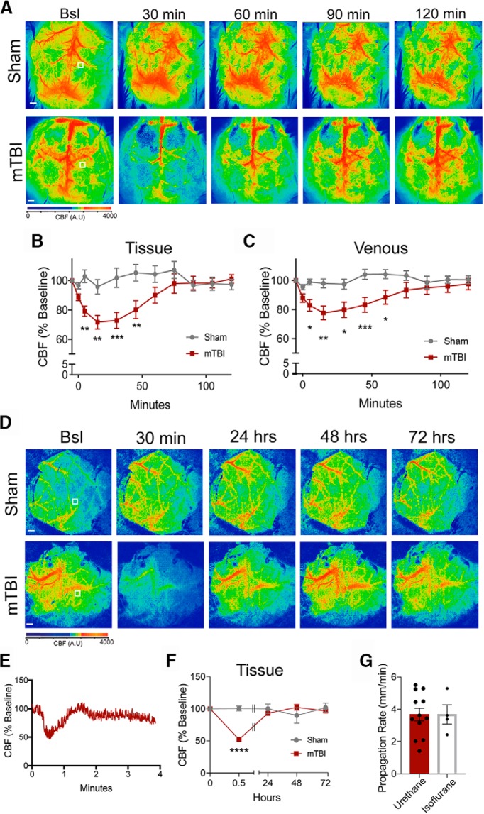
Impact-induced SDs are associated with long-term oligemia. Representative LSCI images of CBF from sham and mTBI animals (A). Representative ROIs indicate the location of repeated measures of CBF in the tissue. CBF was quantified over the 120-min period and plotted over time for sham and mTBI animals in both tissue (B) and venous (C) regions. Modified cranial windows were generated to allow for repeated measures of CBF immediately after the impact and for subsequent days. Representative images before the impact, 30 min post-impact, and subsequent days are shown (D). Representative ROIs indicate where the CBF was quantified. Animals were anesthetized with isoflurane rather than urethane. Representative trace of the hemodynamic responses that are associated with the propagating SD in the presence of isoflurane anesthesia (E). CBF was quantified and normalized to pre-impact baseline. Using LSCI were able to confirm the SD and the peak reduction of CBF and subsequent days following the impact (F). The propagation rate was also quantified in the presence of isoflurane anesthesia (G). Scale bars = 500 μm.

