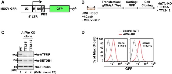
Figure EV1. Establishment and characterization of Atf7ip KO mESCs (related to Fig 1).

-
AThe structure of MSCV‐GFP reporter construct. PBS: primer binding site.
-
BEstablishment of Atf7ip KO cell lines by CRISPR/Cas9 technology.
-
CConfirmation of a complete loss of ATF7IP protein and comparable expression of SETDB1 in the parental WT and established Atf7ip KO cell lines, TT#2‐15 and TT#2‐12, by WB analysis.
-
DFlow cytometric analysis shows that Atf7ip KO cell lines increase the expression of MSCV‐GFP reporter.
Source data are available online for this figure.
