-
A
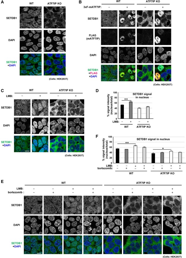
ATF7IP KO HEK293T cells show endogenous SETDB1 cytoplasmic localization phenotype. Endogenous SETDB1 in HEK293T cells shows cytoplasm + nuclear localization profile. Scale bar: 10 μm.
-
B
Over‐production of exogenous mouse SETDB1 induces endogenous SETDB1 nuclear localization in both WT and ATF7IP KO HEK293T cells. The representative data of two independent experiments is shown. Scale bar: 10 μm.
-
C
LMB treatment enhances SETDB1 nuclear accumulation in WT HEK293T cells, but no significant impact for ATF7IP KO HEK293T cells. Cells were treated with 10 ng/ml of LMB for 5 h and then analyzed with immunofluorescent staining. The representative data of three independent experiments are shown. Scale bar: 10 μm.
-
D
SETDB1 signal in the nucleus shown in (C) was calculated. The mean from three independent experiments is shown as a bar graph ± SEM; n = 3. Over 50 cells were analyzed per sample per experiment. **P < 0.01 by Student's t‐test (two‐sided test).
-
E
Five hours of proteasome inhibitor bortezomib treatment does not have much impact on SETDB1 relative nuclear accumulation in ATF7IP KO HEK293T cells. The representative data of three independent experiments are shown. Scale bar: 10 μm.
-
F
SETDB1 signal in the nucleus shown in (E) was calculated. The mean from three independent experiments is shown as a bar graph ± SEM; n = 3. Over 50 cells were analyzed per sample per experiment. Statistics comparison was only shown between without and with drug treatment in WT and ATF7IP KO cells. *P < 0.05 and **P < 0.01 by Dunnett's method.

