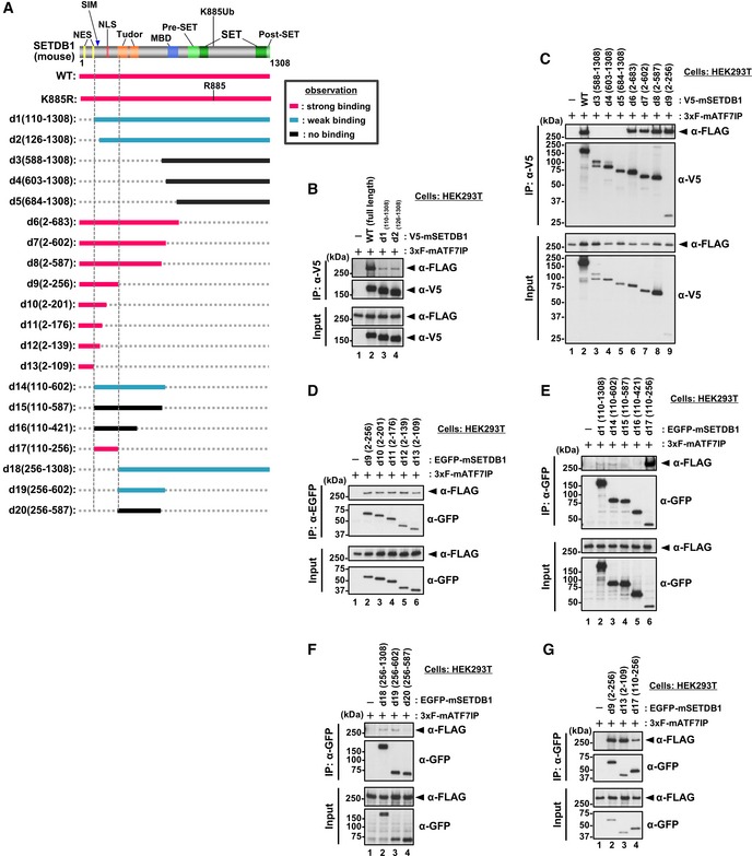
Figure EV4. Mapping analyses for the identification of ATF7IP‐interacting region of SETDB1 (related to Fig 4).

-
ASummary of the co‐IP experiments. After the transfection, V5‐SETDB1 was immune‐purified with anti‐V5 antibody and used for WB analysis. Location of estimated NES and NLS motifs is shown 23.
-
B–GHEK293T cells were co‐transfected with 3xFLAG‐ATF7IP and V5‐tagged or EGFP‐fused SETDB1 WT or deletion mutants as shown in (A). After IP with anti‐V5 antibody (B and C) or anti‐GFP antibody (D–G), the bound ATF7IP was detected by WB analysis. The residues 2–109 (d13) of SETDB1 are mostly sufficient for the interaction with ATF7IP (G).
Source data are available online for this figure.
