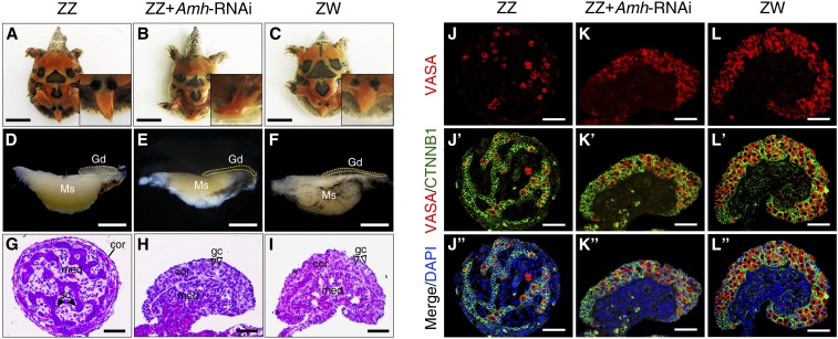
Figure 3.
Feminization of ZZ embryos following Amh knockdown in ovo. (A–C) Morphology of tails from control ZZ, ZZ + Amh-RNAi, and control ZW P. sinensis of stage 27. Bar, 1 cm. (D–F) Representative images of the gonad-mesonephros complexes (GMCs) from control ZZ, ZZ + Amh-RNAi, and control ZW embryos of stage 25. The ZZ gonads with Amh knockdown became elongated and flat compared to control ZZ gonads. Gonads were outlined by yellow dotted lines. Bar, 1 mm. (G–I) H&E staining of gonadal sections from control ZZ, ZZ + Amh-RNAi, and control ZW embryos of stage 25. The ZZ gonads with Amh knockdown appeared thickened outer cortex and degenerated testis cord in medullary region, similar to control ZW gonads. The white dotted lines showed the separation between cortical and medullar regions. Bar, 50 μm. (J–L’’) VASA and CTNNB1 immunostaining of gonadal sections from control ZZ, ZZ + Amh-RNAi and control ZW embryos of stage 25. A female-typical distribution pattern of germ cells was observed in Amh-deficient ZZ gonads. Bar, 50 μm. cor, cortical region; gc, germ cells; Gd, gonad; H&E, hematoxylin and eosin; med, medullary region; Ms, mesonephros; RNAi, RNA interference; sc, Sertoli cell.

