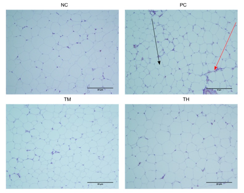
Figure 6.
F4/80 stained immune-histochemical images of white adipose tissue. The black arrow indicates an adipocyte, while the red arrow indicates the brown-stained crown-like structure of the site of macrophage infiltration. For PC, n = 4 samples, and for NC, TM, and TH, n = 4 samples had slides made.

