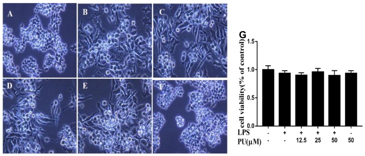
Figure 2.
Photograph of RAW264.7 cells were pre-treated with various concentration of PU for 1 h and treated with LPS for an additional 24 h under optical microscopy (magnification x400). (A) Control; (B) LPS treatment; (C–E) LPS and PU (12.5, 25, and 50 µM) treatment; (F) PU treatment; and (G) cell viability of PU supplementation with LPS-induced RAW264.7 macrophages. The RAW264.7 cells were pre-treated with various concentrations of PU for 1 h and LPS for an additional 24 h. The cell viability was determined by MTT assay, as described in Materials and Methods. The data are presented as means ± SD (n = 3). (**, p < 0.01 vs. control group).

