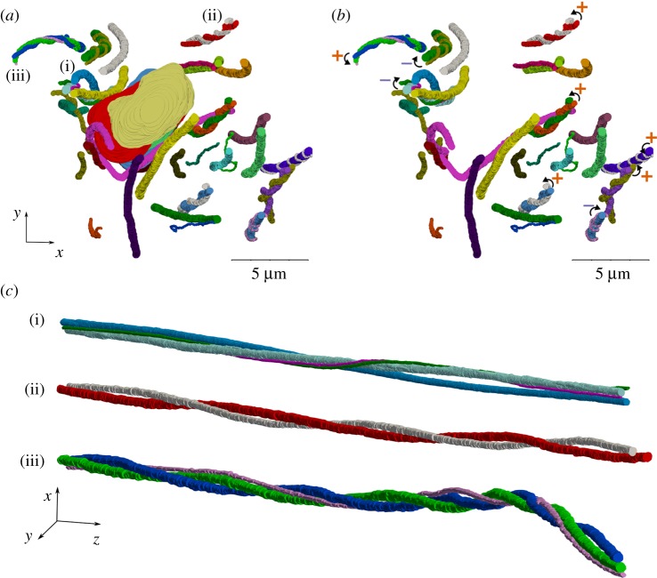
Figure 4.
Many helical structures were observed in the axial view of the fibrils that are evident in the figures (a) with and (b) without the cell nuclei. The helical fibril groups have both right (marked with +ccw curve) and left-handed (marked with – cw curve) twist. (c) Three examples of the groups of twisting fibrils with pitch ranging from 22 to 86 µm. (Online version in colour.)

