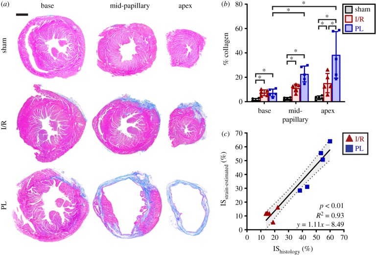
Figure 7.
Histological analysis of collagen content and infarct size. (a) Histology images of mouse left ventricles, obtained 28 days post-surgery and stained with MTC, revealed subepicardial scarring in the I/R group and transmural infarcts in the PL group. Muscle fibres are stained red while collagen-rich scars are stained blue. (b) Bar graphs showing per cent collagen highlighted spatial variations in collagen content relative to infarct location. (c) Strain-estimated infarct size strongly correlated to infarct size measured from the histological midline length approach. Data are shown as mean ± standard deviation (*p < 0.05). Scale bar: 1 mm. (Online version in colour.)

