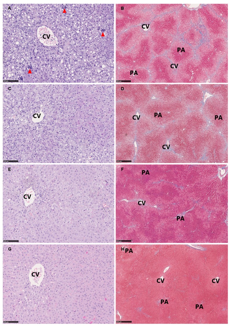
Figure 3.
Representative histological images at study termination (Week 28) of hepatic tissue from: HF (A,B); HF+ (C,D); LF (E,F); and LF+ (G,H). (A,C,E,G) Hematoxylin and eosin stain, scale bar 100 µm. (B,D,F,H) Masson’s trichrome stain (fibrosis stained blue), scale bar 500 µm. Classical pathological lesions of NASH include: steatosis distributed as macro and micro vesicular steatosis, lobular inflammation as inflammatory clusters (red arrowheads) and ballooning hepatocytes (not clearly identified in the current magnification). Advanced and bridging fibrosis is seen in (B,D) (fibrosis in blue). CV, central vein; PA, portal area.

