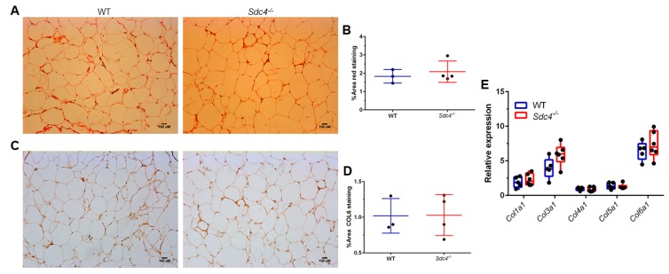
Figure 3.
Sdc4 deficiency does not affect collagen levels in gonadal WATs isolated from female mice fed an HDF for 14 weeks. (A,B) Representative images of picrosirius red staining (Panel A) with quantification (Panel B). (C,D) Representative images of immunohistochemical staining for Collagen VI, alpha 1 (COL6) (Panel C) with quantification (Panel D). In panels B and D, data represent means for n = 3–4 animals, with five not overlapping images taken per animal. Error bars represent standard errors. (E) Gene expression levels were measured by qPCR using mRNA isolated from gWAT. Box and whiskers plots denote individual data points separated by a line representing the group median. Each individual value is plotted as a dot superimposed on the boxplots. Transcript levels of each target gene were normalized to Hprt, Actb, and Tbp.

