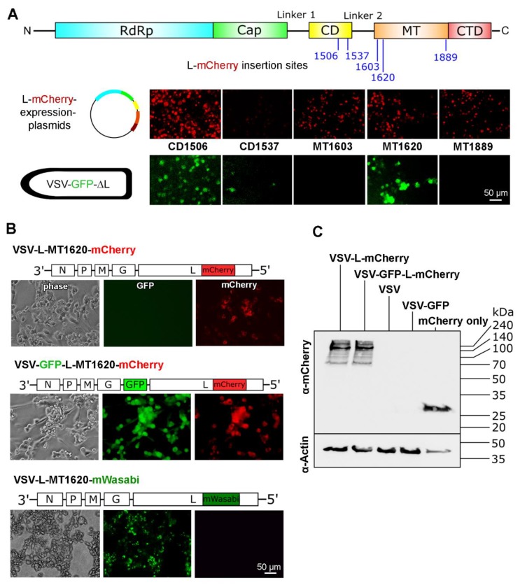
Figure 2.
Insertion of mCherry at position MT1620 is compatible with VSV replication. (A) Top: VSV L-protein domain scheme with insertion sites. Middle: Images of 293T cells transfected with five different L-mCherry expression plasmids. The corresponding insertion sites are labeled with the domain abbreviation followed by amino acid number. Red indicates L-mCherry expression. Bottom: GFP signal after inoculation with VSV-GFP-ΔL at a multiplicity of infection (MOI) of 10 depicted from the same micrograph frames as above. Green fluorescence indicates functional L-mCherry fusion proteins and polymerase activity. Scale bar 50 µm. (B) Fluorescence and phase contrast images of VSV-L-mCherry and VSV-GFP-L-mCherry 24 h after infection of BHK-21 cells: Virus genome schemata are displayed above the fluorescence images. Scale bar 50 µm. (C) Immunoblot against mCherry under reducing conditions on 12% polyacrylamide gel: β-actin was used as loading control. VSV, VSV-GFP, VSV-L-mCherry, and VSV-GFP-L-mCherry infected BHK-21 cells 8 h after infection were used to prepare lysates.

