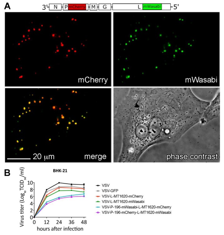
Figure 6.
Attenuated double P- and L-insertion viruses display co-localizing fluorescent signals: (A) Assessment of putative P- and L-protein interaction in a representative micrograph of BHK-21 cells infected with VSV-P-mCherry-L-mWasabi at MOI of 10 at 10 h post infection. (B) Viral replication kinetics of parental VSV, VSV-GFP, VSV-L-MT1620-mCherry, VSV-L-MT1620-mWasabi, VSV-P-mWasabi-L-mCherry, and VSV-P-mCherry-L-mWasabi compared in BHK-21 at 37 °C in a multistep replication kinetics (MOI 0.1) at time points 12, 24, 36, and 48 h after infection. Titers were quantified using TCID50. Data shown as mean (SD) from two independent samples.

