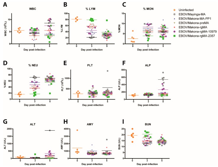
Figure 3.
Hematology and blood biochemistry in mice who were infected with MA-EBOV. Groups of mice (n = 6) were infected intraperitoneally with 10 PFU of either EBOV/Mayinga-MA, EBOV/Makona-MA-PP1, EBOV/Makona-preMA, EBOV/Makona-rgMA, or the two single mutant viruses EBOV/Makona-rgMA-10979 or EBOV/Makona-rgMA-2357. Blood samples were collected on days 3 and 5 post-infection and (A) total white blood cell count (WBC), (B) % lymphocytes (%LYM), (C) % monocytes (%MON), (D) % neutrophils (%NEU), (E) platelets (PLT), (F) alkaline phosphatase (ALP), (G) alanine aminotransferase (ALT), (H) amylase (AMY), and (I) blood urea nitrogen (BUN) were measured. Blood was also collected on day 0 from a group of uninfected BALB/c mice (n = 6) to establish baseline levels for all the parameters.

