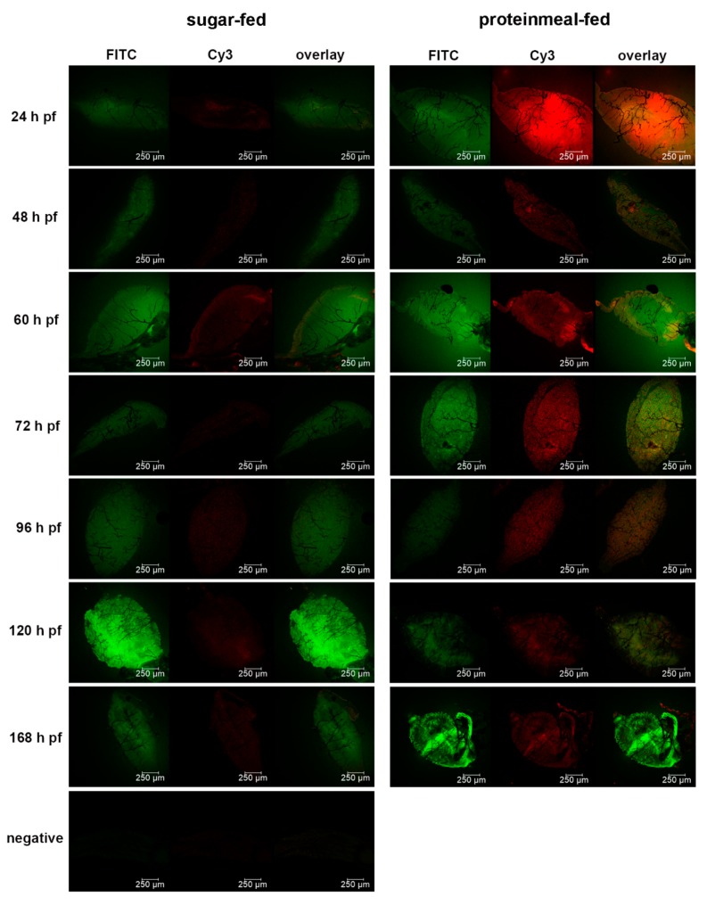
Figure 4.
Detection of 5 nm and 50 nm gold-nanoparticles in midguts obtained from sugar-fed and proteinmeal-fed (20% BSA solution) mosquitoes at various time points post-feeding (pf) by fluorescence microscopy. Dissected midguts were soaked for 2 h in a suspension containing 5 nm (1.99 × 10⁹ nanoparticles/mL, labeled with FITC) and 50 nm (1.99 × 10⁹ nanoparticles/mL, labeled with Cy3) gold-nanoparticles before mounting and viewing. Midgut samples were viewed under an inverted spectral confocal microscope (TCP SP8 MP, Leica Microsystems) thereby maintaining the same imaging parameters for each sample.

