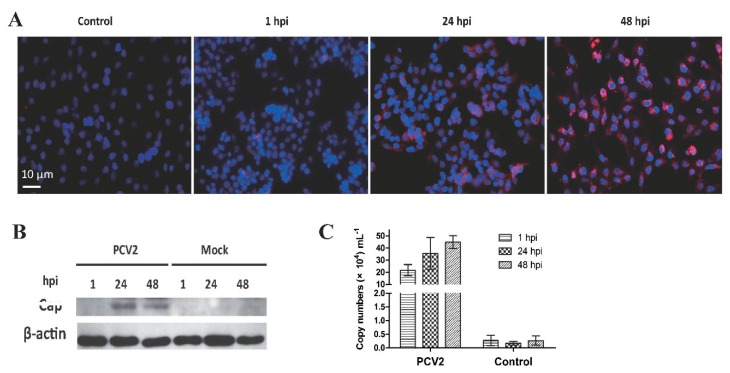
Figure 1.
Porcine circovirus type 2 (PCV2) infection in porcine alveolar macrophages (PAMs). (A) PAMs were infected with PCV2b and processed for immunofluorescence assay at 1, 24, and 48 hpi. PCV2b was visualized by the ODs-psdAb probe against the Cap protein (red), the nucleus was stained with DAPI (blue). (B) Cap protein in PCV2b-infected PAMs was also detected by Western blot assay based on Cap protein (VLPs) immunized porcine serum. (C) PCV2 copy numbers were analyzed by qPCR at the time-points indicated. Results are mean ± SE of three independent experiments.

