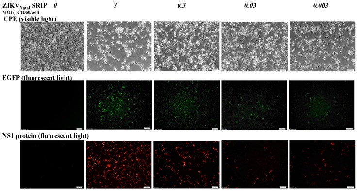
Figure 5.
Infectivity of ZIKV Natal RGN SRIPs in prM-E-expressing packaging cells. The cells were infected with ZIKV Natal RGN SRIPs at MOIs of 3, 0.3, 0.03, and 0.003. The cytopathic effect (top) and the EGFP reporter (middle) in SRIP-infected cells were photographed using light and fluorescence microscopy 72 h post-infection. In addition, infected cells were washed, fixed, and stained by anti-NS1 antibodies and Alexa Fluor 546-conjugated secondary antibodies (bottom). Finally, cell imaging was taken by immunofluorescence microscopy. Scale bars, 100 μm for CPE and EGFP images (top and middle) and 200 μm for the NS1 image.

