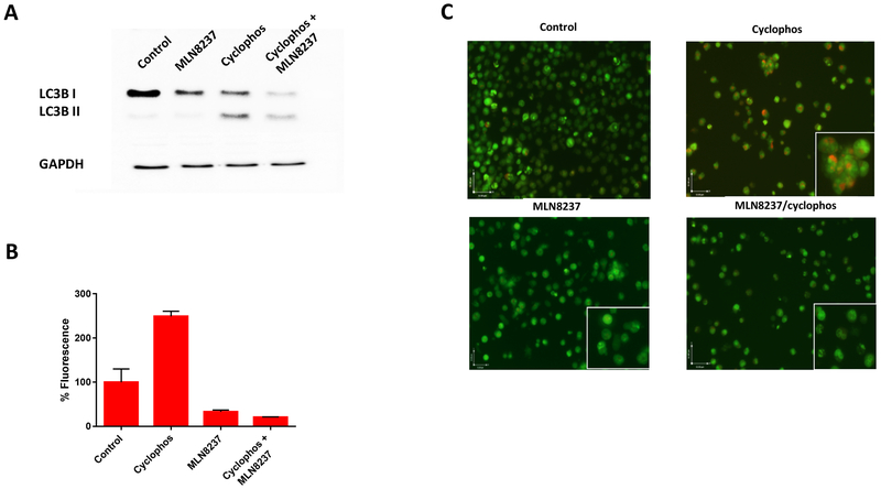
Figure 6.
AURKA inhibition reverses autophagy. (a) LC3B-II levels by immunoblotting in chemoresistant (Raji) cells treated with PBS (negative control), cyclophosphamide, MLN8237, or combination of cyclophosphamide and MLN8237, or etoposide (positive control) at 48 hours. (b) % Fluorescence of autophagy influx measured by flow cytometry. (c) Representative photomicrograph (400 X) of autophagy levels in Raji cells stained with after treatment acridine orange staining with PBS (negative control), cyclophosphamide, MLN8237, or combination of cyclophosphamide and MLN8237. The accumulation of acidic vesicular organelles, which emit bright red/orange fluorescence correspond to autophagy activity.

