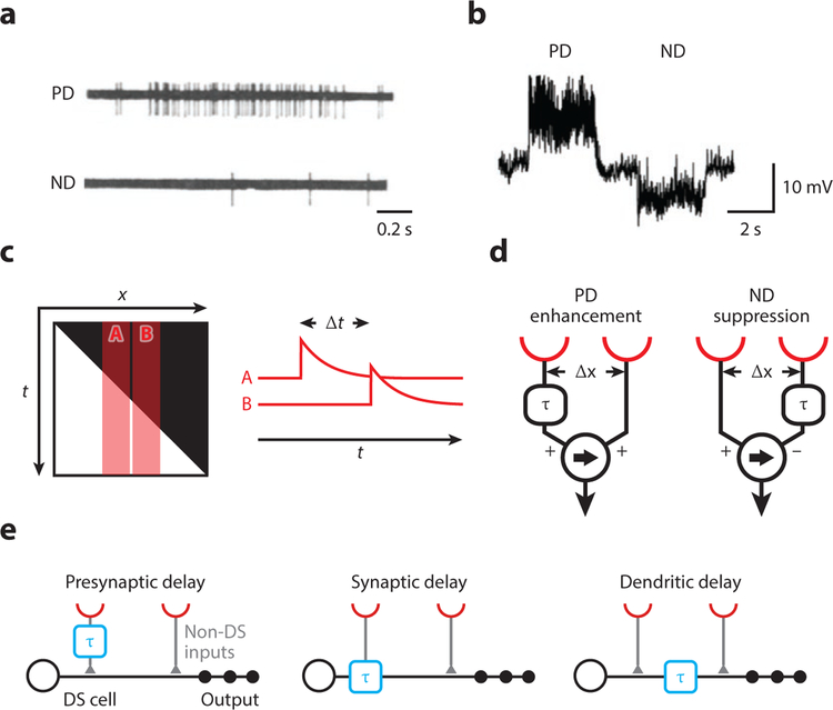
Figure 1. Cells and models for visual motion detection.
(a) Spiking response of a direction-selective unit in the rabbit retina to a moving light spot. Panel adapted with permission from Barlow and Levick (1965).
(b) Response of a lobula plate tangential cell in Drosophila to a grating moving in opposite directions. Panel adapted with permission from Joesch et al. (2008).
(c) The brightness profile of a light edge moving to the right shown in space-time (x-t). A and B represent the receptive fields of two adjacent photoreceptors, which are activated sequentially with a delay Δt. The traces A and B depict a high-pass-filtered version of a signal, highlighting illumination changes.
(d) Two alternative motion detector models. Both generate direction-selective responses by a delay-and-compare mechanism but differ in their nonlinear integration. In the Hassenstein-Reichardt model on the left, a delayed signal (denoted by τ) enhances a direct signal, for instance by a multiplication. In the Barlow-Levick model on the right, a delayed signal suppresses a direct signal, for instance by a division. In both models, the arrow indicates the preferred direction.
(e) Potential cellular implementations of local motion detection (not mutually exclusive). Spatially offset signals are conveyed through different cell types or different synaptic receptors with different dynamics (left and middle, respectively). Temporal delays might also arise by dendritic filtering (right). Note that “presynaptic delay” could be generated by an arbitrary mechanism in the upstream circuit. Abbreviations: DS, direction-selective; ND, null-direction; PD, preferred-direction.

