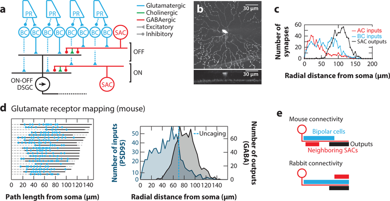
Figure 2. Visual motion detection in the mammalian retina.
(a) Schematic of the mammalian retinal circuit elements for direction selectivity. Photoreceptor signals are split into an ON and an OFF pathway at the level of bipolar cells via different glutamate receptors. ON and OFF bipolar cells synapse individually onto direction-selective ON and OFF SACs, respectively, and jointly onto ON-OFF DSGCs. DSGC receive additional asymmetric GABAergic and symmetric cholinergic input from SACs. Note that only selected aspects of the connectivity are captured in this simplified schematic.
(b) A z-projection of an ON SAC filled with a fluorescent dye showing the radial dendrites and stratification.
(c) Distribution of inputs and outputs determined by serial electron microscopy reconstructions of mouse ON SACs as well as their pre- and postsynaptic partners. Panel adapted with permission from Ding et al. (2016).
(d) (left) Distributions of glutamatergic inputs on SAC dendrites determined using glutamate uncaging. Each row is a different SAC dendrite, the solid line is the mapped region of the dendrite, and blue dots are glutamatergic synapses as revealed by glutamate uncaging. (right) Comparison of glutamatergic inputs determined by labeling SACs with the postsynaptic marker for glutamatergic synapses, PSD95-YFP (blue) and outputs determined using serial electron microscopy reconstructions. The average location of the most distal synapse measured with uncaging is represented by the dotted line. Panel adapted with permission from Vlasits et al. (2016).
(e) Schematic of dendritic locations of inputs (blue, bipolar cells; red, neighboring SACs) and outputs (black) in mouse and rabbit SACs. Abbreviations: AC, amacrine cell; BC, bipolar cell; DSGC, direction-selective ganglion cell; EM, electron microscopy; PR, photoreceptor; SAC, starburst amacrine cell.

